Patients: 60 post-THR patients (29 and 1 did not complete) and post-TKR patients (30)
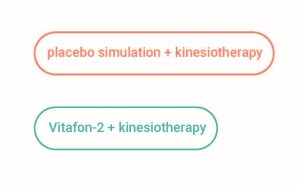
Study design: 1Post – TKR patients: 19 – Vitafon-2 + kinesiotherapy; 11 – placebo simulation + kinesiotherapy
Post – THR patients: 20 – Vitafon-2 + kinesiotherapy; 9 – placebo simulation + kinesiotherapy
Study duration: About 1 week (procedures on the on the second, third and fourth day post-surgery)
Medical device: Vitafon-2
Research methods: Leg circumference in the groin for post-THR patients and just above the patella for post-TKR patients, pain intensity by VAS scale, active knee flexion and extension and active hip flexion, abduction, adduction and rotation using a goniometer, monitoring the haematoma
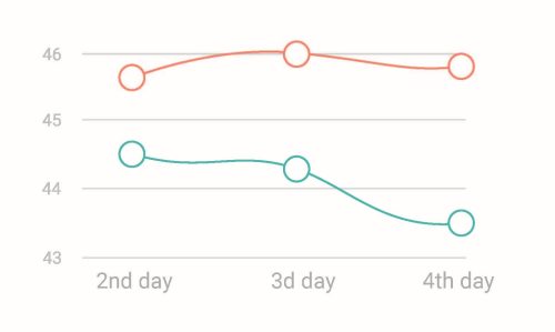
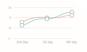
Dynamics in knee circumference (post-TKR).
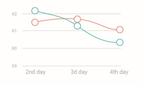
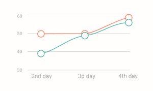
Dynamics in groin circumference (post-THR).
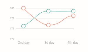
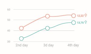
Dynamics in knee extension angle among post-TKR patients.
Dynamics of internal rotation angle among post-TKR patients.
Dymamics in knee flexion angle among post-TKR patients.
Dynamics of external rotation angle among post-TKR patients.
Patients: 20 patients diagnosed with Dupuytren’s disease and operated on for contracture (mainly the second and third stage)
Study design: 10 patients – parallel laser (CTL 1106MX) and viroacoustic treatment with Vitafon,
10 patients – only Vitafon
Study duration: 10-14 days after the surgery when the sutures were removed
Medical device: Vitafon-T
Research methods: Measuring the range of motion in metacarpal-phalangeal joints (MCP), proximal interphalangeal joints (PIP) and proximal interphalangeal joints (DIP) expressed in centimetres (distance between the finger pulp and the styloid line during maximal flexion); estimation of pain by Laitinen Questionnaire and Visual Analog Scale (VAS)
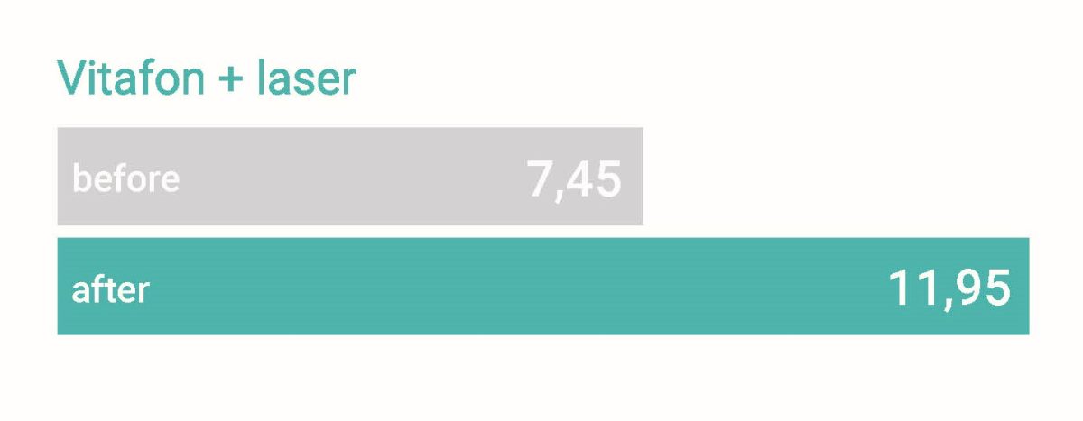
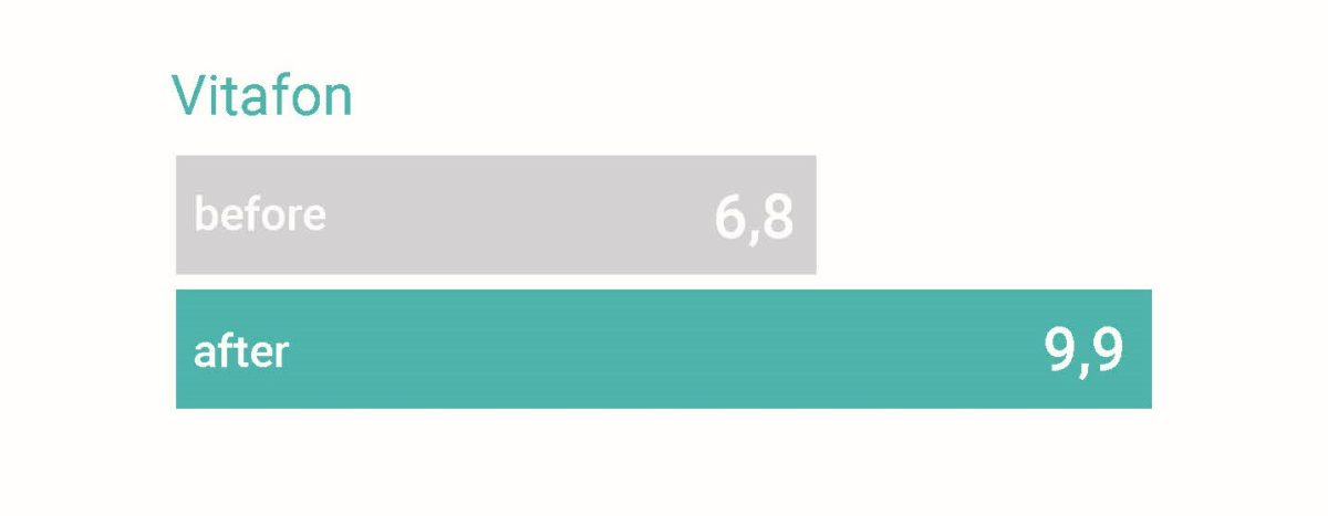
Dynamics in active total range motion before and after therapy (cm).
The range of flexion both in MCP and PIP joints: increased in Vitafon + laser group average by 3.4 cm and in Vitafon group by 2.2 cm.
The extension range both in MCP and PIP joints improved on average by 1.1 cm in group Vitafon + laser and 0.9 cm in group Vitafon.
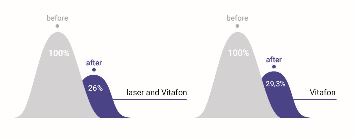
Decrease in pain in % (VAS).
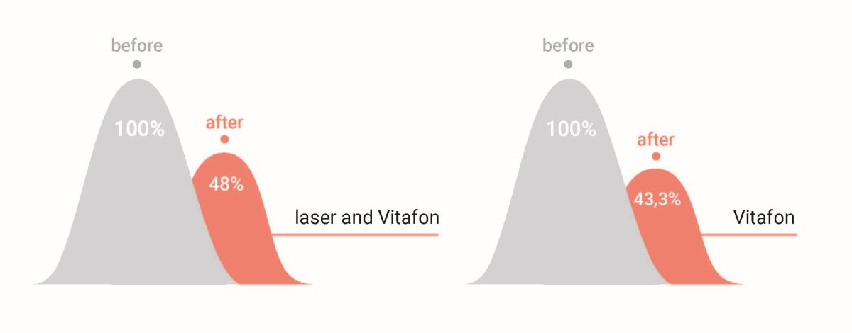
Dynamics in freqeuency of pain in %.
Patients: Two experimental studies with rabbits (16 and 30),then clinical:
I. 71 patients with fractures of the tubular bones of the hand
II. 30 patients after surgery of the closed fractures of the metacarpal bones of the hand
Research design: Two experimental studies with rabbits, then clinical:
I. 35 – Vitafon group +standard treatment, 36 – control group (only standard treatment)
II. 35 – Vitafon group, 36 – control group (magnetotherapy)
Study duration: I. II.
Device (model): Vitafon (I) and Vitafon-T (II)
Research methods: Experimental study: X-ray, X-ray densitometric and morphological and morphometric study
Clinical study: densitometry (Lunar Prodigy Advance), rheovasography, time of disability, functional results on scales DASH (Disabilities of the Arm, Shoulder and Hand) and Kollontai scale summing the scores for a) range of finger movements, b) grips and hand strength c) consolidation dynamics based on radiographs, d) labor recovery period.
Experimental studies with rabbits have shown that in Vitafon group the swelling regresses faster, osteogenesis is more active that is already seen on the 6th day of observation.
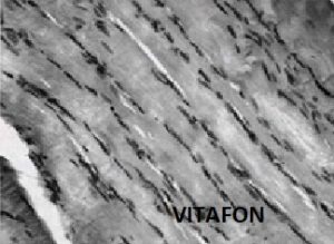
Control group (rabbits)
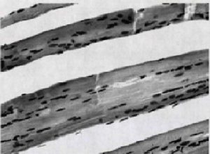
In the soft tissues outside the fracture zone, pronounced swelling was observed cervical tissue and focal myocyte dystrophy. cm in group Vitafon.
VITAFON group (rabbits)
In the soft tissues outside the damage zone, the swelling was significantly less pronounced than in the control at a similar observation period, which was expressed by the tight fit of dystrophically altered muscle fibers to each other.
On the 6th day in Vitafon group signs of soft tissue swelling and inflammatory cellular reaction of soft tissue damage have already disappeared (while in control group – not)
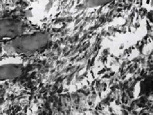
Control group (rabbits)
In the soft tissues in damaged zone persistent chaotic arrangement of fibroblasts in granulations filling tissue detritus. Hematoxylin and eosin staining.
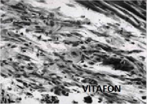
VITAFON group (rabbits)
In the soft tissues of the damage zones, granulations replacing tissue detritus of animals in the experimental group looked more mature than in the control. In them, bundles of unidirectional elongated fibroblasts were located parallel to each other
On the 6th day in Vitafon group in the area of soft tissue damage, more mature granulations are fixed.
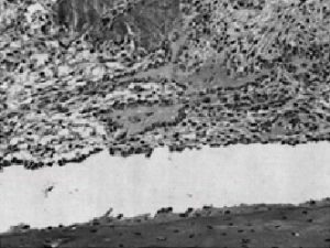
Control group (rabbits)
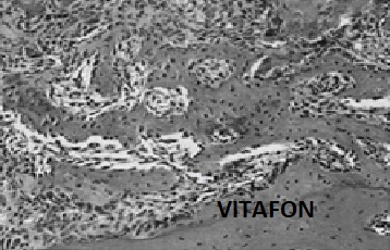
In the bone tissue, the formed defects were filled with cellular tissue detritus with acellular bone fragments in it. In the granulations of the periosteal and endosteal surfaces of the edges of the distal defect, signs of osteogenesis were found in the form of the formation of small osteoid structures.
VITAFON group (rabbits)
In all three formed bone defects of animals in the experimental group, signs of osteogenesis were observed, which was most pronounced in the distal defect: its lumen along the periosteal surface was completely blocked by granulation tissue with a network of delicate osteoid structures.
On the 21st day after modeling the defects in the Vitafon group rabbits, the bone regenerate that filled the defect looked more mature than in the control. Its compaction was observed with restoration of the structure of the cortical plate and the contours of th bone marrow canal, which did not occur in the animals of the control group. In the area of the distal foramen of the animals in the experimental group, a complete restoration of the structure of the cortical plate was observed, while in the animals of the control group all three defects were easily detected by structural changes in the bone tissue.
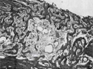
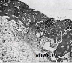
Control group (rabbits)
IThe distal defect is covered by unevenly calcified, merging bone beams. Periosteal layers of newly formed bone are subject to osteoclastic resorption.
VITAFON group (rabbits)
Compacting bone regenerates filling defects. In one of the defects there is a patch of unreplaced fibrous tissue with resorption granulomas
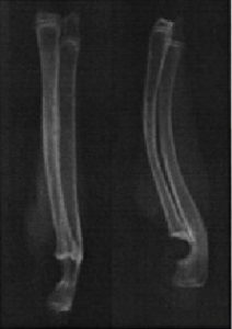
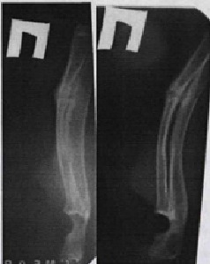
According to the radiographs taken on the 16th and 21 animals, however, in the Vitafon group the callus has been more pronounced with massive periosteal growths area of the defect of the radial bone of the forearms.
Control group (rabbits)
VITAFON group (rabbits)
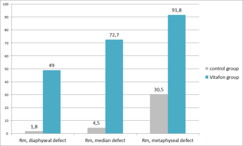
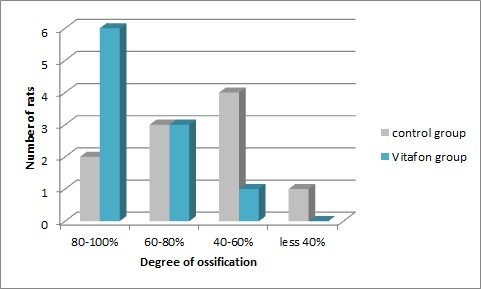
Based on the results of morphometric analysis, histological signs of greater maturity of the regenerate were identified in animals of the Vitafon group. The statistical significance of these differences with control group for all defect localizations was confirmed using the Mann-Whitney test.
Results of morphometric analysis (Rm%). Mann-hitney test
Clinical trial:
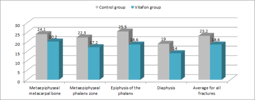
The duration of consolidation in the Vitafon group (18.6±3.2 days) is shorter than in the control group (23.2±2.2 days) by an average of 5.2 days.
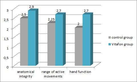
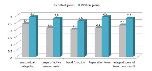
Assessment of hand functionality (scores) by Kollontai scale
The degree of restoration of the anatomical structure of the bone was higher by 0.4 points in the Vitafon group, and the range of active movements and hand function were higher by 0.5 and 0.7 points, respectively. The results obtained convincingly prove that against the background of vibroacoustic influence, bone callus forms faster, after removing the plaster splint, the limitation in the range of active movements is less pronounced and hand function is restored faster.
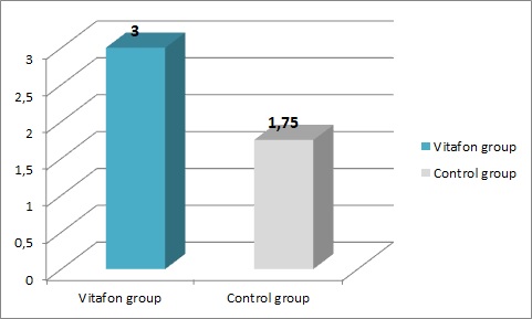
Results of assessment of hand function on the 24th day
Vibroacoustic influence has a qualitative effect on the regenerative process, optimizing osteogenesis. In the Vitafon group, the ratio of the density of the fracture zone to the density of the entire hand was significantly higher. Thus, when analyzing radiographs using the “fracture” program, an uneven distribution of optical density in favor of the damaged zone was revealed (both in the experiment in experimental animals and in the clinic in the main group).
Patients: I. Experimental: 29 laboratory rats
II. 112 people with fractures of the tubular bones of the hand (osteoepiphysiolysis SHII, epiphyseal fracture SHIII, avulsion fracture, diaphysis fracture) aged 8 to 17 years
epiphyseal fracture SHIII, avulsion fracture, diaphysis fracture) aged 8 to 17 years
Research design: I. Experimental.
a. 9 rats underwent osteoperforation of the upper third of the femur. Then they were divided into 3 groups: 3 rats received therapy on the 1st mode of the Vitafon device, 3 rats on the 2nd Vitafon mode, and 3 rats without Vitafon therapy.
b. In 20 rats, a femoral bone defect was created by perforating the proximal metadiaphysis of the femur. 10 rats – with Vitafon, 10 rats – without Vitafon.
II. Group 62 – Vitafon, 50 – control – without Vitafon
Study duration: 4 years of clinical practice
Medical device: Vitafon-2 (vibroacoustic and infared stimulation)
Research methods: I. Experimental: morphological analysis, histology.
II. Clinical: assessment of range of motion in the corners using an inclinometer, dynamometry, X-ray examination on the day of injury, on the 7th day, on the 14th day, on the 21st day, on the 28th day, assessment of hand functions using the method of Kollontai
Yu.Yu. (1983) as modified by Davydov Yu.I. (2001), namely summing the scores for a) range of finger movements, b) grips and hand strength c) consolidation dynamics based on radiographs, d) labor recovery period.
According to the results of an experimental study in a group of rats receiving Vitafon therapy:
in the early stages:
• less pronounced swelling of the soft tissues was observed (in the majority of animals in the Vitafon group, the swelling disappeared completely on the 5th day, in the control group only by the 14th day)
• more active cellular response to damage, expressed in lymphoplasmacytic infiltration
• the presence of active proliferation of fibroblastic elements, which was not observed in the control group.
in later stages:
• more active formation of osteoid structures at the edges of the bone defect.
• faster recovery of motor activity of the injured limb.
Morphological analysis showed that due to the effect of Vitafon on the area of the perforation hole, the reparative ability of the red one marrow is enhanced, endosteal bone formation is stimulated, the primary formation of bone beams in the area of the callus, i.e. optimal conditions are created for the formation of primary callus.
According to the results of a clinical study in children in the Vitafon group:
• swelling went away faster (by the time the plaster splint was removed in children in the main group, swelling remained in only 1 patient out of 62, while in patients in the control group – in 10 out of 50).
• pain was relieved faster (pain syndrome in children in the Vitafon group was relieved on average 5 days earlier than in children in the control group).
• the range of motion in the joints of the hand in children from the Vitafon group was fully restored on average 2.5 days earlier than in the control group. Limitation of range of motion after removal of the plaster splint was observed in only 26 patients in the main group, while in all patients in the control group.
• hand strength after removal of the plaster splint, according to dynamometry data, was restored 2 days earlier in children of the Vitafon group than in the control group.
The main indicator of the effectiveness of treatment was the duration of fracture consolidation. The degree of consolidation was determined by X-ray films. The duration of consolidation in the Vitafon group (18.6 days) is less than in the control group (23.2 days) by an average of 4.6 days. The effect on the duration of consolidation is most pronounced in cases of epiphyseal fractures (the difference is 6.9 days). Improved blood circulation has a particularly beneficial effect on repair processes in intra-articular fractures.
The maximum positive effect of Vitafon was manifested in a faster recovery period in the main group – 22.8 ± 3.0 days, in the control group 32.3 ± 4.0 days, which is manifested in a faster return of the child to school, sports, music and other sections, the difference was 9.5 days. During the recovery period, the volume of active movements significantly increases, restoration of hand function occurs 9.5 days (31.0%) earlier compared to traditional patient management
Patients: 114 patients with tubular fractures hand bones after fixing bones
Research design: 59 Vitafon-group, 56 – control group
Study duration: 2 years
Medical device: Vitafon
Research methods: rg-граммы и оценивалась консолидация перелома, по оригинальному способу, разработанному на отделении, применением программы «перелом» Оценка функциональных результатов производилась по методике Колонтай Ю.Ю. в модификации Давыдова Ю.В.
In the main group, the use of vibroacoustic effects reduced the consolidation time by an average of a week, which made it possible to remove metal structures earlier, stop immobilization and begin physical therapy.
The period of incapacity for work in the Vitafon group was reduced by an average of 10 days.
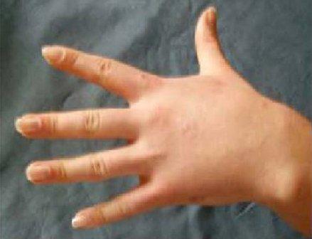
On the photo: clinical example: a patient on the 28th day after closed reduction and osteosynthesis of the 5th
metacarpal bone (after a closed fracture): full restoration of function.
Rehabilitation of children with scarring consequences of burns
Patients: 50 children with scarring consequences of burns
Research design: Comparison to usual practice
Medical device: Vitafon used to enhance the effect of Contractube x gel
Research methods: infrared sensing and clinical observation
According to the study, the full evolution of hypertrophic scars in children ended by 8–9 months after surgical or spontaneous restoration of the skin, which should be considered as a positive result. In all patients, an acceleration in the organization of scars was noted, which was clinically expressed in the absence of itching, a decrease in the height of the scar relative to healthy skin, and an increase in its elasticity and mobility relative to the underlying tissues.